Home » Without Label » Anatomy Of Chest - Rotation of 3D skeleton.ribs,chest,anatomy,human,medical ... - The superior thoracic aperture found superiorly and the inferior thoracic aperture.
Anatomy Of Chest - Rotation of 3D skeleton.ribs,chest,anatomy,human,medical ... - The superior thoracic aperture found superiorly and the inferior thoracic aperture.
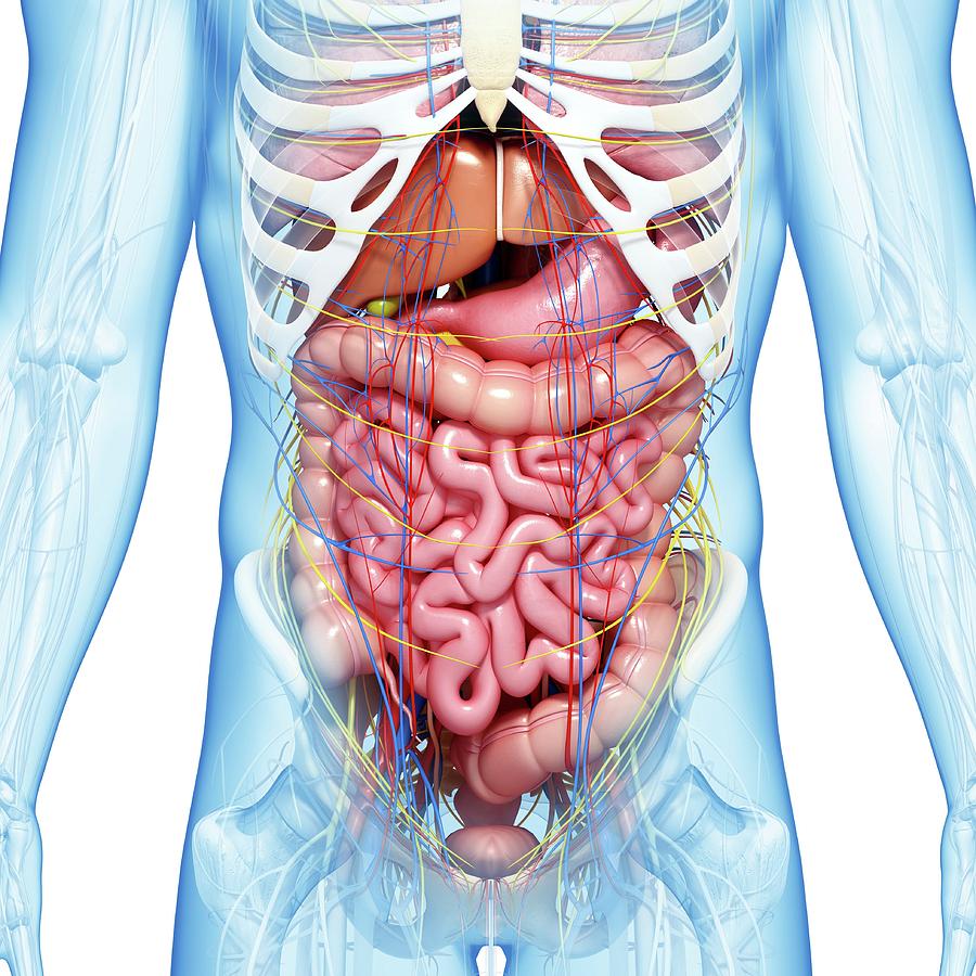
Anatomy Of Chest - Rotation of 3D skeleton.ribs,chest,anatomy,human,medical ... - The superior thoracic aperture found superiorly and the inferior thoracic aperture.. It is enclosed by the ribs, the vertebral column, and the sternum, or breastbone, and is separated from the abdominal cavity (the body's largest hollow space) by a muscular and membranous partition, the diaphragm. Here, we break down the anatomy of your chest muscles. 31 anatomy of the female breast syllabus p. Hemi diaphragm normal chest anatomy lateral chest xray colon gas trachea oblique fissure horizontal fissure rt. The circulatory system does most of its work.
Large, complex chest wall defects can be some of the most challenging problems a reconstructive surgeon must face, but successful outcomes may be reliably achieved by adhering to basic principles of adequate debridement followed by. An overview of the anatomy visible in a transverse computed axial tomographical image of the thorax (and part of the abdomen) performed with intravenous cont. Browse 6,407 chest anatomy stock photos and images available, or search for human anatomy to find more great stock photos and pictures. The superior thoracic aperture found superiorly and the inferior thoracic aperture. 30 lines of the thoracic wall syllabus p.
Anatomy of a Chest of Drawers - FineWoodworking from s3.amazonaws.com Anatomy of right side chest pain. This chapter is an abbreviated review of thoracic anatomy as seen on chest radiographs and computed tomography (ct) of the chest. The chest is made up primarily of two muscles: A line is drawn from anterior surface of the body of 6th thoracic vertebrae passing through the apex of the heart up to anterior lower most part of diaphragm. Anatomy of the female human body 12 photos of the anatomy of the female human body. An overview of the anatomy visible in a transverse computed axial tomographical image of the thorax (and part of the abdomen) performed with intravenous cont. Basic thoracic anatomy and physiology an understanding of thoracic imaging requires knowledge of the anatomy being imaged, as described in this chapter, as well as the imaging techniques applied to the thorax, covered in chapter 2. Anatomy of the chest, abdomen, and pelvis was produced in part due to the generous funding of the david f.
This page provides an overview of the chest muscle group.
The chest wall is comprised of skin, fat, muscles, and the thoracic skeleton. This page provides an overview of the chest muscle group. 12 cm (5 in) in length, 8 cm (3.5 in) wide, and 6 cm (2.5 in) in thickness. Anatomy of the thorax, heart, abdomen and pelvis recommended text gray's anatomy for students. It provides protection to vital organs (eg, heart and major vessels, lungs, liver) and provides stability for movement. The chest is the area of origin for many of the body's systems as it houses organs such as the heart, esophagus, trachea, lungs, and thoracic diaphragm. An overview of the anatomy visible in a transverse computed axial tomographical image of the thorax (and part of the abdomen) performed with intravenous cont. 30 lines of the thoracic wall syllabus p. See human chest anatomy stock video clips. You will also find the xiphoid process, 10th rib, the apex of the heart, the coronary vein of the heart. Anatomy of the thorax, heart, abdomen and pelvis recommended text gray's anatomy for students. Here, we break down the anatomy of your chest muscles. The chest anatomy includes the pectoralis major, pectoralis minor and the serratus anterior.
It spreads out like a fan and covers the rib cage like an armor plate. The chest wall is comprised of skin, fat, muscles, and the thoracic skeleton. #anatomy of the chest and stomach. The thorax has two major openings: Thoracic wall the first step in understanding thorax anatomy is to find out its boundaries.
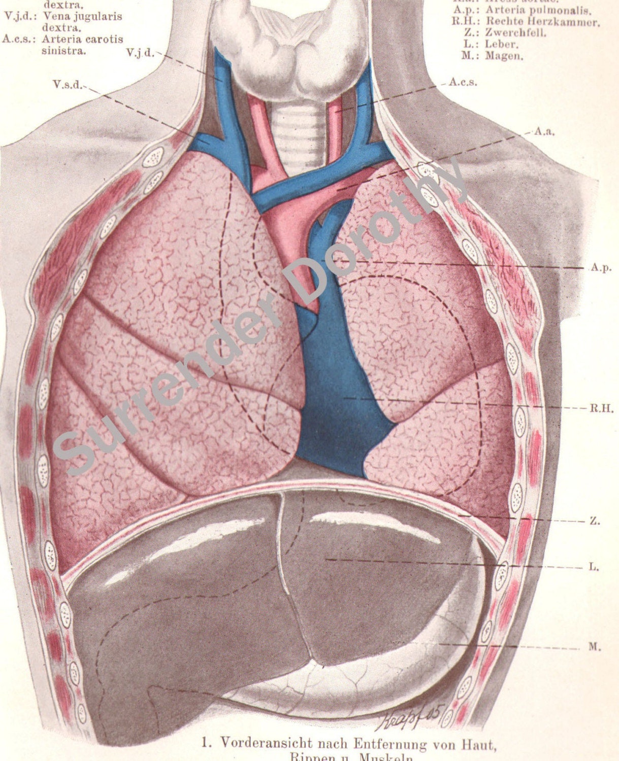
Abdominal Anatomy Photograph by Pixologicstudio/science ... from images.fineartamerica.com Anatomy of the female human body 12 photos of the anatomy of the female human body. In insects, crustaceans, and the extinct trilobites, the thorax is one of the three main divisions of the creature's body, each of which is in turn composed of multiple segments. The chest anatomy includes the pectoralis major, pectoralis minor and the serratus anterior. Swensen fund for innovation in teaching. Sternocleidomastoid muscle clavicle and ribs anatomy muscle anatomy chest sternocleidomastoid ribs anatomy chest muscles anatomy thorax rib muscles chest muscles chest anatomy illustration. Principal functions are the protection of internal viscera and an expandable cylinder facilitating variable gas flow into the lungs. Table 1.1 lists the major anatomic structures within the thorax that are discussed. In this image, you will find common carotid arteries, internal jugular vein, subclavian artery, subclavian vein, heart, right lung, 6th rib, diaphragm, costal cartilage in it.
Chest muscles anatomy (1) pectoralis major muscle.
Swensen fund for innovation in teaching. The superior thoracic aperture found superiorly and the inferior thoracic aperture. The chest or thorax is the region between the neck and diaphragm that encloses organs, such as the heart, lungs, esophagus, trachea, and thoracic diaphragm. A line is drawn from anterior surface of the body of 6th thoracic vertebrae passing through the apex of the heart up to anterior lower most part of diaphragm. Table 1.1 lists the major anatomic structures within the thorax that are discussed. Related posts of anatomy of the chest area anatomy of the female human body. The myotomes elongate and invade the mesoderm of the wall of the embryonic thoracic and abdominal cavities. Sternocleidomastoid muscle clavicle and ribs anatomy muscle anatomy chest sternocleidomastoid ribs anatomy chest muscles anatomy thorax rib muscles chest muscles chest anatomy illustration. Anatomy of the thorax, heart, abdomen and pelvis recommended text gray's anatomy for students. The chest or thorax region of the upper body has a number of important organs that reside within it that may present with chest pain if they become compromised in. 12 photos of the anatomy of the chest and stomach. Chest bone, ribs, lung, heart, xiphoid process, sternum anatomy. Principal functions are the protection of internal viscera and an expandable cylinder facilitating variable gas flow into the lungs.
Table 1.1 lists the major anatomic structures within the thorax that are discussed. Large, complex chest wall defects can be some of the most challenging problems a reconstructive surgeon must face, but successful outcomes may be reliably achieved by adhering to basic principles of adequate debridement followed by. A typical heart is approximately the size of your fist: Anatomy of the chest, abdomen, and pelvis was produced in part due to the generous funding of the david f. The chest or thorax region of the upper body has a number of important organs that reside within it that may present with chest pain if they become compromised in.
Vintage Human Anatomy Lungs Chest 1906 Medical Chart | Etsy from i.etsystatic.com The muscles of the chest develop from the somites found in the mesoderm. These myotomes divide into the epimere and the hypomere. The myotomes elongate and invade the mesoderm of the wall of the embryonic thoracic and abdominal cavities. Learn about each of these muscles, their locations, functional anatomy and exercises for them. Knowledge of the anatomy of the whole cylinder (ribs, sternum, vertebra, diap … A typical heart is approximately the size of your fist: The thorax or chest is a part of the anatomy of humans, mammals, other tetrapod animals located between the neck and the abdomen. 4 innervation of the breast blood supply of the breast syllabus p.
The muscles of the chest develop from the somites found in the mesoderm.
12 photos of the anatomy of the chest and stomach. Here, we break down the anatomy of your chest muscles. You will also find the xiphoid process, 10th rib, the apex of the heart, the coronary vein of the heart. Anatomy of the thorax, heart, abdomen and pelvis recommended text gray's anatomy for students. Chest a man's chest — like the rest of his body — is covered with skin that has two layers. The pectoralis major and the pectoralis minor, known collectively as your pecs. (1) the pectoralis major, and (2) the pectoralis minor. Learn about each of these muscles, their locations, functional anatomy and exercises for them. The chest or thorax region of the upper body has a number of important organs that reside within it that may present with chest pain if they become compromised in. The chest wall is comprised of skin, fat, muscles, and the thoracic skeleton. The muscles of the chest develop from the somites found in the mesoderm. The anatomic illustrations are presented as… Plus, how to target each to make them bigger and stronger.